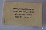
2. ABC-M8 Chemical Agent Detector Paper (M8 Paper)
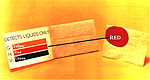
M8 paper (Figure 6) is used to test liquid substances for the presence of nerve agents and blister agents.[25] It is similar to the litmus (pH) paper that is found in almost any laboratory in that a test result is indicated in both types of paper by a change in color. The difference is that M8 paper is specifically designed to react to nerve agents and blister agents in liquid form.[26] It cannot be used to test vapors for the presence of CWAs. Originally included in the older generation detection and sampling kits, M8 paper was incorporated into the M256 kit without modification.[27] It is also issued to individuals as a separate piece of CWA detection equipment. Each M256 kit comes with one booklet of M8 paper that contains 25 sheets. Unused M8 paper is tan in color and has three sensitive indicator dyes suspended within the paper. M8 paper responds (changes color) within 30 seconds of exposure to liquid G and V nerve agents and H or L blister agents. Each of these CWAs has a different level of acidity (pH). The detector dyes react to the different pH levels by changing to one of three colors. The color yellow indicates the presence of a G nerve agent; the color green indicates a V nerve agent. Exposure to either H or L blister agents produces a red color (see Figure 7). M8 paper does not have to be completely saturated with a liquid chemical agent to produce a test result. It reacts to a liquid measure as small as 0.02 milliliter (a drop or two).[28]
3. Sensitivity to Chemical Warfare Agents
The amount of a CWA absorbed through the skin or inhaled over a period of time is referred to as the dose that an individual has received. Although each person responds in a predictable way (e.g., develops burns and blisters on the skin, develops blurred vision, or loses consciousness, etc.) after becoming exposed to a CWA, the time it takes for symptoms to first appear may vary slightly from individual to individual. In addition, the dose that an individual must absorb to cause a reaction can also vary slightly from individual to individual. Dosage levels have been established that will predictably cause a reaction in a defined percentage of a population. The two dosage designations used in this paper are the effective dose (that which causes an effect in a defined percentage of a population) and lethal dose (that which causes death in a defined percentage of the population). The effective dose has been established for most CWAs, while the lethal dose is generally known for all CWAs. In addition, the effective dose may differ from the lethal dose for an agent depending on its form, liquid or vapor. Effective dose and lethal dose are further explained in Table 1.[29][30]
Table 1. Explanation of effective dose and lethal dose[31]
| Chemical Warfare Agent | Dosage | Explanation |
| Liquid Form | Effective Dose: ED50 | The letters E or L differentiate between effective and lethal doses and the subscript number depicts the applicable percentage of the population. In this case, the number 50 represents 50 percent of the population; 25 percent of the population would be identified as ED25 or LD25. |
| Lethal Dose: LD50 | ||
| Vapor Form | Effective Dose: ECt50 | The letters E or L differentiate between effective and lethal doses and the subscript number depicts the applicable percentage of the population (the number 50 represents 50% of the population). The letter C represents the concentration or the amount of agent that is suspended in one cubic meter (m3) of air (e.g., 10 mg, 20 mg, 50 mg, etc.); the letter t represents the period of exposure in minutes. A lethal dose expressed as: LCt50 5 mg x minute/m3 means that 50% of the population would die if they received a dose that consisted of a concentration of 5 milligrams of agent for one minute (5 multiplied by 1) in one cubic meter (m3) of space. |
To prevent injury and death to personnel, the M256 kit must be capable of detecting the presence of a CWA at a concentration level that is lower than a concentration level that could injure personnel. This assists commanders, or other responsible individuals, to determine if a CWA's concentration is below a casualty-producing level and that it is safe to remove MOPP gear. Table 2 provides a comparison between the known effective and lethal doses of CWAs and the M256 kit’s minimum effective detection capability, while Table 3 provides a comparison of the capabilities of other CWA detectors used by the US military. The purpose for including these tables is twofold. The first reason is to show, by comparison, the effectiveness of the M256 kit in detecting CWAs at much lower levels than CWAs are known to cause injuries and death (Table 2). The second reason is to show that the M256 is able to detect CWAs at much lower concentration levels than other detection devices used by US military personnel (Table 3).
Table 2. Comparison of effective and lethal doses of chemical warfare agents with the M256 Detection Kit’s minimum detection capability|
|
Agent |
Liquid |
M8 Paper |
Vapor/Aerosol |
M256 |
||
| Effective Dose (ED50) |
Lethal Dose LD50 (skin) |
Effective Dose (ECt50) |
Lethal Dose (LCt50) |
||||
| N
|
GA |
*[33] |
1 g/70-kg man[34] |
.02 ml | 2 – 3 mg†[35] |
400 mg[34] |
.005 mg |
GB |
*[33] |
1.7 g/70-kg man[34] |
.02 ml | 3 mg†[33] |
100 mg[34] |
.005 mg |
|
GD |
*[33] |
350 mg/70-kg man[34] |
.02 ml | 1 mg†[33] |
50 mg[34] |
.005 mg |
|
GF |
*[33] |
30 mg[33] |
.02 ml | 1 mg[33] |
*[33] |
.005 mg |
|
VX |
1 mg[33] |
10 mg/70-kg man[34] |
.02 ml | <1 mg†[33] |
10 mg[34] |
.02 mg |
|
| B |
H |
10 ug[34] |
7 g/70-kg man[34] |
.02 ml | 10 mg[34] |
1,500 mg[34] |
2 mg |
HD |
10 ug[33] |
7g/70-kg man[34] |
.02 ml | 10 mg[34] |
1,500 mg[34] |
2 mg | |
| CX | *[33] |
*[33] |
n/a |
300 mg[34] |
3,200 mg[34] |
3 mg | |
| L | 15 ug‡[33] |
40 – 50 mg/70-kg man[34] |
.02 ml | Eye: 30 mg Skin:~200 mg[34] |
1,500 mg[34] |
9 mg |
|
| B L O O D |
AC | *[33] |
100 mg[34] |
n/a |
~1500 mg[33] |
2,500 - 5,000 mg[34] | 9 mg |
| CK | *[33] | *[33] | n/a | *[33] | 11,000 mg[34] | 8 mg |
|
* Indicates that a dose has not been reliably estimated. † The approximate amount that would cause pinpointing of the eye (miosis) when the eye comes into direct contact with the chemical agent as opposed to exposure through inhalation (respiratory) or being absorbed through the skin (percutaneous). ‡ The approximate amount that would cause redness or inflammation of the skin (erythema). |
Table 3. Comparison of US detection devices
| Detection Device | Chemical Warfare Agent | Minimum Detectable Amount | ||
| M8A1 Alarm[36] | Nerve Agents | G – series | .1 - .20 mg | |
| VX | .4 mg | |||
| CAM[37] |
Nerve Agents | G – series VX | .1 mg | |
| Blister Agents | HD and HN | |||
| M18A2 Kit[38] | Nerve Agents | GB and VX | .1 mg (Enzyme method) | |
| GB | 1.0 mg (Adsorption method) | |||
| Blood Agent | AC | 8.0 mg | ||
| Blister Agents | HD L |
.5 mg 10.0 mg |
||
| Choking Agent | CG | 12.0 mg | ||
| MM-1 Mobile Mass Spectrometer (Fox vehicle equipment)[39] | Nerve Agent | GB | 62 mg | |
| Blood Agent | CK | 46 mg | ||
| Choking Agent | CG | 115 mg | ||
| M256 Kit[40] | M8 Paper | Vapor-Sampler | ||
| Nerve Agent | GA | .02 ml | .005 mg | |
| GB | .02 ml | .005 mg | ||
| GD | .02 ml | .005 mg | ||
| GF | .02 ml | .005 mg | ||
| VX | .02 ml | .02 mg | ||
| Blister Agent | H | .02 ml | 2 mg | |
| HD | .02 ml | 2 mg | ||
| CX | n/a | 3 mg | ||
| L | .02 ml | 9 mg | ||
| Blood Agent | AC | n/a | 9 mg | |
| CK | n/a | 8 mg | ||
4. Instructions for Using the M256 Kit
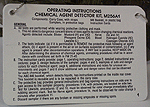
Instructions, warnings, and advisory information for operating the M256 kit test components are contained in the operator’s manual. For ease of use under battlefield conditions, several variations of the instructions have been prepared with battlefield conditions in mind. A condensed version of the vapor-sampler’s instructions and advisory information are printed on plastic laminated instruction cards that accompany each kit (Figure 8). Although the instruction cards do not provide the same level of detail as the operator’s manual, they are sufficient to guide an operator through the test from start to finish as well as to interpret test results. The cards provide step-by-step instructions for using the sampler. They also provide diagrams to aid the operator in identifying the sampler’s components, and color charts for interpreting test results. Instructions are printed on both sides of the card so the operator need only flip a card up to move from the bottom of one page to the top of the next. In addition to the instruction cards, a bare bones version of the instructions and advisories are printed on both sides of the vapor-sampler’s protective envelope (Figure 9).
Figure 8. M256 kit instructions
Figure 9. Vapor-Sampler's instructions on side 1 of the protective envelope
There are no step-by-step instructions for using M8 detection paper other than those contained in the operator’s manual. A color chart used to interpret test results is located on the inside front cover of each tablet of M8 paper (Figure 7).
A. How the M256 Vapor-Sampler Works
Each type of CWA has its own unique chemical make-up that produces a predictable colored reaction when mixed with certain other chemicals. When using the vapor-sampler to test vapors and aerosols, the colored reaction indicates whether a CWA is present. It takes 15 to 20 minutes to detect the presence of a blood agent, nerve agent, or blister agent using the M256 test kit, which includes a 10-minute period when the vapor-sampler is exposed to the gas or vapor in question.[41] After the test is complete, the operator compares the test spots with the positive (danger) and negative (safe) test result examples printed both on the instruction cards and on the vapor-sampler body.
1. Blood Agent Test and Blister Agent Test
When performed successfully, both the blister agent test and blood agent test will produce a uniquely colored result. If either agent is present at a concentration level that is equal to or greater than the M256 kit’s minimum detectable concentration level, the agent-specific color will develop on the test spot.
To perform the blister agent test, the appropriate glass ampoule marked with the number "3" (Figure 10) is crushed.[42] This releases a solution known as 4-(4-Nitrobenzyl) Pyridine (DB3) and mercuric cyanide in methanol[43] that flows downs its channel to moisten the square-shaped blister agent test spot. The solution reacts with both H series blister agents and CX.[44] Next, the moistened test spot is heated using the attached chemical heater for two minutes. After heating, the blister agent test-spot is exposed to the suspect vapor for 10 minutes. During the exposure period, the vapor-sampler can be laid down or held by the hinged protective strip to expose the vapor-sampler to the suspect vapor. After 10 minutes, the blister agent test-spot is heated a second time for one minute. After heating, the glass ampoule marked with the number "5" is crushed to apply a potassium carbonate solution[45] to the test spot. If an H blister agent is present, the potassium carbonate causes the spot to turn blue-purple. If CX is present, the spot will turn pink or red.[46]
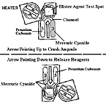
Figure 10. Releasing blister agent test reagents
Blood agents (AC and CK) are detected using the round-shaped test spot that has been impregnated with barbituric acid. To initiate the blood agent test, the operator crushes the two glass ampoules in the center of the vapor-sampler and marked with the number "3" (Figure 11). This releases test reagents — sodium hypochlorite and a solution of 4-benzyl pyridine in 2-methoxy ethanol.[47] If the blood agent AC is present, the sodium hypochloride converts it to the blood agent CK. CK reacts with barbituric acid and 4-benzyl pyridine to form the pink-red or blue color.[48]
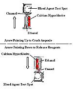
Figure 11. Releasing blood agent test reagents
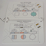
The operator compares the color of the test blister agent and blood agent test spots against the safe observation and danger observation diagrams which are part of the instructions that come with each M256 kit (Figure 12). The operator can also compare the color changes against the safe-danger observation information that is included on the front side of each vapor sampler.
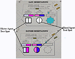
Figure 12. Example of blood agent and blister agent test results
2. Lewisite TestThe lewisite test has two components: a tan colored, crayon-like pellet (containing Milcher’s thioketone[49] and other inert solids) and a paper-rubbing pad. The pellet is contained in a plastic cup molded into the vapor-sampler’s body. The paper-rubbing pad, which is white in color, is located on the underside of the tab (inset in Figure 13). When performed successfully, the lewisite test will also produce a uniquely colored result. If lewisite is present at a concentration level that is equal to or greater than the M256 kit’s minimum detectable concentration level, the agent specific color (olive green) will develop on the rubbing paper.[50]
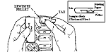
Figure 13. Rubbing the lewisite pellet and paper rubbing pad
To conduct the lewisite test, the paper pad is rubbed against the test pellet (Figure 13). About 10 minutes later, as directed by the M256 kit’s directions, a second rub mark is made on the paper. The two marks are then compared to each other. If the color of the first mark has turned to olive green, lewisite is present. If the first mark remains tan in color, lewisite is not present at a concentration level that would produce injury or casualties (Figure 14).[51]
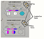
Figure 14. Example of lewisite test results
The nerve agent test is conducted in the same manner as the blood and blister agent tests, i.e., glass ampoules are crushed and the test spot is moistened (Figure 15). Unlike the other chemical agent tests (blister, blood, and lewisite), a change in the color of the nerve agent test spot from clear, or having no color, to blue-green means that no agent is present (Figure 16).[52] The star-shaped test spot is coated with an enzyme (called acetylcholinesterase) which has been extracted from electric eels.[53] A buffer solution from one of the glass ampoules (marked with the number "3") is applied to the test spot to moisten the dried enzyme. After 10 minutes of exposure to the air, indoxyl acetate is applied from a second ampoule (marked with the number "5"). If a nerve agent is not present, a blue color will appear on the test spot. However, if a nerve agent is present, there is no change in color to the test spot.[54]
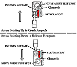
Figure 15. Releasing Nerve Agent Test Reagents
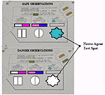
Figure 16. Example of Nerve Agent Test Results
To test liquids for the presence of a chemical agent, a piece of M8 paper is attached to a probe (or similar device) or held in a gloved hand.[55] The paper is then blotted, not rubbed, into the suspect liquid. Rubbing the M8 paper into the suspect liquid may cause abrasions on the paper and cause false positive (red) detection streaks.[56] After the M8 paper is exposed to a liquid, the person conducting the test then compares the color changes on the paper to the color chart inside the cover of the M8 paper tablet: yellow indicates the presence of a G nerve agent; green indicates VX nerve agent; and red indicates either H or L blister agents. If the paper does not change color, CWAs that M8 paper is capable of detecting are not present at the minimum detectable concentration level of the detection paper.[57]
| First Page | Prev Page | Next Page |